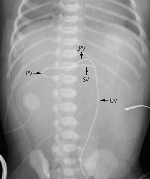


Some complications can occur in a well-positioned catheter. Malpositioning within the portal venous system is associated with portal vein thrombosis. If the umbilical venous catheter reaches the left portal vein but does not continue into the ductus venosus, the catheter can travel left into the more peripheral left portal vein or right, where it can eventually course into the right portal vein or hepatofugally into the main portal vein (or potentially farther into the vessels that merge to form the portal vein: the superior mesenteric and splenic veins). Place the tip of the catheter, held with forceps (see Figure 1) into the lumen of the vessel and gently advance to 5 cm into the vein. left atrium and beyond (through a patent foramen ovale or an atrial septal defect).If the umbilical venous catheter is advanced too far along its intended course, the tip may end up in a number of locations: Alternatively, the catheter may not travel along the intended route (wrong turn). The catheters are inserted by the pediatrician without imaging guidance, and given the small size of infants (especially those requiring umbilical catheters), a small variation in length of catheter can result in significant malpositioning (too long).

Anomalous positioningĪnomalous positioning of the umbilical venous catheters is quite frequent. The tip should lie at the junction of the inferior vena cava with the right atrium. It passes through the umbilicus, umbilical vein, left portal vein, ductus venosus, middle or left hepatic vein, and into the inferior vena cava. Preparation of a complete UVC insertion kit should occur well in advance of a planned home birth and should be complete and readily available for every home. An umbilical venous catheter generally passes directly superiorly and remains relatively anterior in the abdomen.


 0 kommentar(er)
